Sonoanatomy
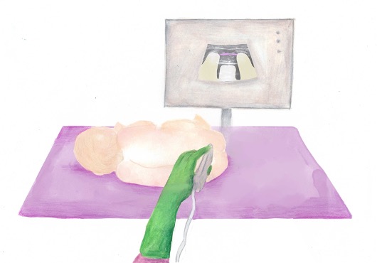
Ultrasound probe selection: linear high frequency probe
Mapping/scout scan: look between the intervertebral spinous processes and laminae:
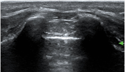
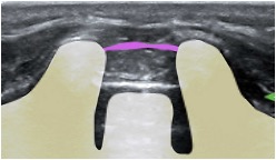
1)Probe position transverse over the sacral cornua (-> frog’s eyes US view)
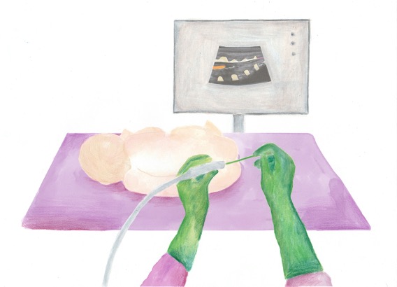
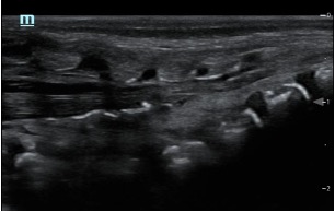
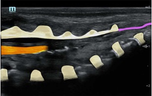
2)Then rotate probe 90 degrees for a longitudinal view
Identify:
•Sacral cornua
•Sacrum
•Sacrococcygeal membrane
•End of the dural sac (hyperechoic (<-> CSF are anechoic (black)
•Caudal epidural space
•Assess the position of the dural sac in relation to the sacrococcygeal membrane
NOTE: Ultrasound can't see through bone; so with increasing age (= increasing angulation of the spinous processes and increasing ossification of the laminae) the size of the echo window will diminish -> you might need to scan paramedian
TIP: use a large linear probe (50mm) – this will allow you to visualise more vertebrae in one image for easier monitoring of needle approach and LA spread
Paediatric Plan A Blocks
About
News
Education
Courses
Research
Resources
Grants
Webinars
ASM
Members
Plan A
Plan A Paeds
Plan A Block Videos
Terms & Conditions
Privacy
ASFRA/RAUK Meeting 2026
13th annual conference of the African Society of Regional Anesthesia & Pain Therapy
Membership Terms and Conditions
Content Creators

